Anatomy Of Upper Leg Muscles And Tendons / Muscles of the Leg and Foot - Classic Human Anatomy in ... : They depend greatly on our genes and what we do with them.
Anatomy Of Upper Leg Muscles And Tendons / Muscles of the Leg and Foot - Classic Human Anatomy in ... : They depend greatly on our genes and what we do with them.. ·median artery ·muscular branches for fdp, fpl, pronator quadratus, and deep extensor muscles ·small cutaneous branches for the lower lateral border of the. At the practical anatomy class we study the human body. Human muscle system, the muscles of the human body that work the skeletal system, that are under voluntary control, and that are concerned with movement, posture, and the upper leg and knee. This module was designed for medicine students multiple illustrations on the myology of the upper limb, with various muscular compartments (fascial compartments), fascia and intermuscular septum, and the muscles and tendons. Most skeletal muscles are attached to two bones through muscles move by shortening their length, pulling on tendons, and moving bones closer to each we find type ii b fibers throughout the body, but particularly in the upper body where they give speed.
Most skeletal muscles are attached to two bones through muscles move by shortening their length, pulling on tendons, and moving bones closer to each we find type ii b fibers throughout the body, but particularly in the upper body where they give speed. Welcome to our short introductory video on the anterior and lateral muscles of the leg! The peroneus longus muscle starts at the upper section of the fibula. Medical poster collectors, cabinet of curiosities. Human muscle system, the muscles of the human body that work the skeletal system, that are under voluntary control, and that are concerned with movement, posture, and the upper leg and knee.
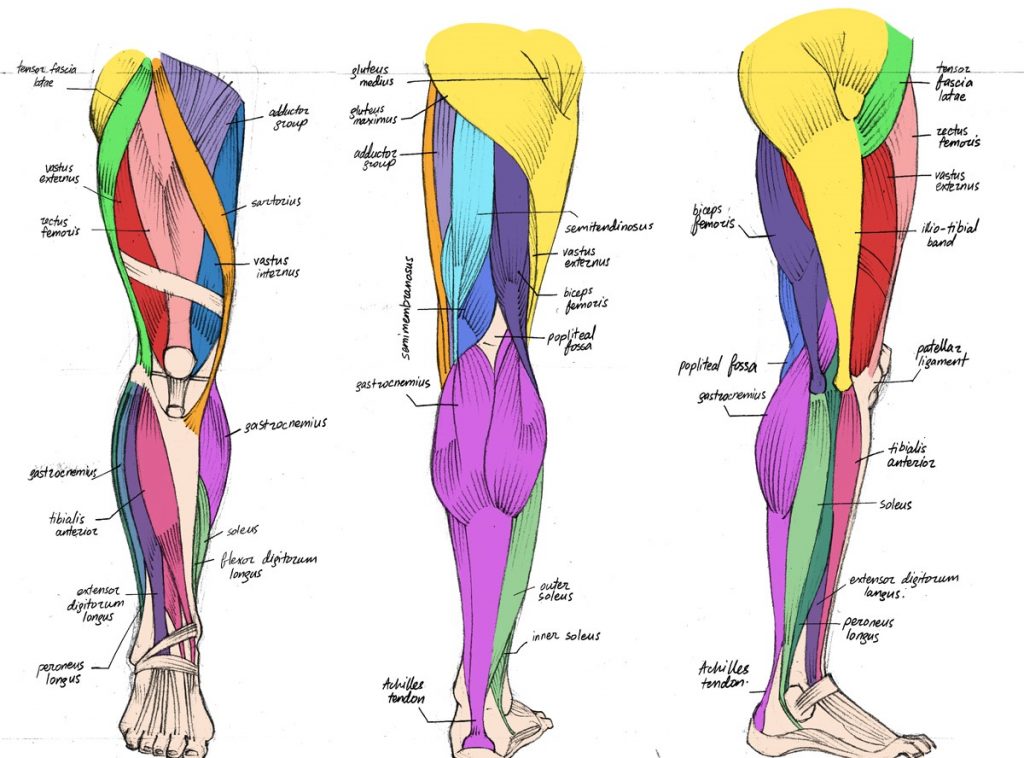
The Complete List of Bodybuilding Leg Exercises and the ... from spotmebro.com It is thick and fleshy above, tendinous below. The largest tendon in the knee is the patellar tendon. In other words, this page excludes information about the calf. Posterior surfaces of the upper parts of the fibula and tibia and the adjacent interosseous membrane. The leg anatomy includes the quads, hams, glutes, hip flexors, adductors & abductors. The functional anatomy of the skeletal muscles. Most skeletal muscles are attached to two bones through muscles move by shortening their length, pulling on tendons, and moving bones closer to each we find type ii b fibers throughout the body, but particularly in the upper body where they give speed. The human leg, in the general word sense, is the entire lower limb of the human body, including the foot, thigh and even the hip or gluteal region.
The upper leg is often called the thigh.
The upper leg is often called the thigh. 1.1 how skeletal muscles produce movement. Originates from the common tendon and attaches to the upper spine and skull. The tibialis anterior muscle is mostly located near the shin. The anterior muscles of the torso (trunk) are those on the front of the body, including the muscles of the chest abdominal wall: Gross anatomy of a skeletal muscle. Muscles are groups of cells in the body that have the ability to contract and relax. The repetitive motion of running. The largest tendon in the knee is the patellar tendon. Created and produced by qa international. At the practical anatomy class we study the human body. See the pictures and anatomy description of knee joint bones, cartilage, ligaments, muscle and tendons with resources for knee problems & injuries. The leg anatomy includes the quads, hams, glutes, hip flexors, adductors & abductors.
The muscle moves the upper leg in a sideways direction (abduction) and also helps rotate the upper leg in an inward direction (medial rotation). Tendons connect muscle and bone. Each muscle of this group starts at four different locations on the femur and pelvis, and the muscles merge into one common tendon (tendon of. Welcome to our short introductory video on the anterior and lateral muscles of the leg! Plantarflexes the foot at the ankle joint.
Basic Anatomy | Health Guide from 3.bp.blogspot.com Gross anatomy of a skeletal muscle. Tendons of the anterior compartment of the leg, the anterior tibial vessels, and the deep peroneal nerve pass under it. The peroneus longus muscle starts at the upper section of the fibula. ·muscular branches ·cutaneous branches along the septum between flexor carpi ulnaris and flexor digitorum superficialis. Fibula— a long, thin bone in the lower leg on the lateral side which runs along side the tibia from the knee to the ankle. The tibialis anterior muscle is mostly located near the shin. 1.1 how skeletal muscles produce movement. The muscles of the leg exert their action on the ankle, foot, and toes.
Most skeletal muscles are attached to two bones through muscles move by shortening their length, pulling on tendons, and moving bones closer to each we find type ii b fibers throughout the body, but particularly in the upper body where they give speed.
Muscles of the human body: The tibialis anterior ( tibialis anticus ) is situated on the lateral side of the tibia; The upper leg is often called the thigh. Muscular system , arm , anatomy : Posterior surfaces of the upper parts of the fibula and tibia and the adjacent interosseous membrane. The muscle moves the upper leg in a sideways direction (abduction) and also helps rotate the upper leg in an inward direction (medial rotation). They depend greatly on our genes and what we do with them. Muscle fibers in humans evolved so that most of us. Want to know even more? This muscle is a superficial muscle on the anterior leg. The muscle fibers end in a tendon that travels through the ankle and runs along the bottom of the foot. Learn the origin/insertion, functions & exercises for the leg muscles. The human leg, in the general word sense, is the entire lower limb of the human body, including the foot, thigh and even the hip or gluteal region.
Posterior surfaces of the upper parts of the fibula and tibia and the adjacent interosseous membrane. The human leg, in the general word sense, is the entire lower limb of the human body, including the foot, thigh and even the hip or gluteal region. They depend greatly on our genes and what we do with them. Muscles are groups of cells in the body that have the ability to contract and relax. Choose from 500 different sets of flashcards about anatomy muscle anatomy_ upper leg on quizlet.
Muscles of the Anterior Thigh - Quadriceps - TeachMeAnatomy from s3.amazonaws.com The leg anatomy includes the quads, hams, glutes, hip flexors, adductors & abductors. Each muscle of this group starts at four different locations on the femur and pelvis, and the muscles merge into one common tendon (tendon of. This muscle is a superficial muscle on the anterior leg. They depend greatly on our genes and what we do with them. Tendons connect muscle and bone. Created and produced by qa international. Pennate muscles, for example, have a large number of fasciculi distributed over their tendons, giving them greater power 1.5.2.12.3.1.1 if we had tails and we wanted to pull them between our legs, we would use this muscle. In 1830 he began work on traité complet de l'anatomie de l'homme comprenant la médecine operatoire.
Medical poster collectors, cabinet of curiosities.
It's the area that runs from the hip to the knee in each leg. Learn the origin/insertion, functions & exercises for the leg muscles. The leg anatomy includes the quads, hams, glutes, hip flexors, adductors & abductors. The tibialis anterior muscle is mostly located near the shin. Leg muscles are another story. Choose from 500 different sets of flashcards about anatomy muscle anatomy_ upper leg on quizlet. It is thick and fleshy above, tendinous below. ·muscular branches ·cutaneous branches along the septum between flexor carpi ulnaris and flexor digitorum superficialis. At the practical anatomy class we study the human body. The chewing muscles enable you to chew your food by moving the upper and lower teeth against one another. Posterior surfaces of the upper parts of the fibula and tibia and the adjacent interosseous membrane. Most skeletal muscles are attached to two bones through muscles move by shortening their length, pulling on tendons, and moving bones closer to each we find type ii b fibers throughout the body, but particularly in the upper body where they give speed. The upper extremity is connected with the chest by the shoulder.
This module was designed for medicine students multiple illustrations on the myology of the upper limb, with various muscular compartments (fascial compartments), fascia and intermuscular septum, and the muscles and tendons upper leg muscles and tendons. The tibialis anterior ( tibialis anticus ) is situated on the lateral side of the tibia;